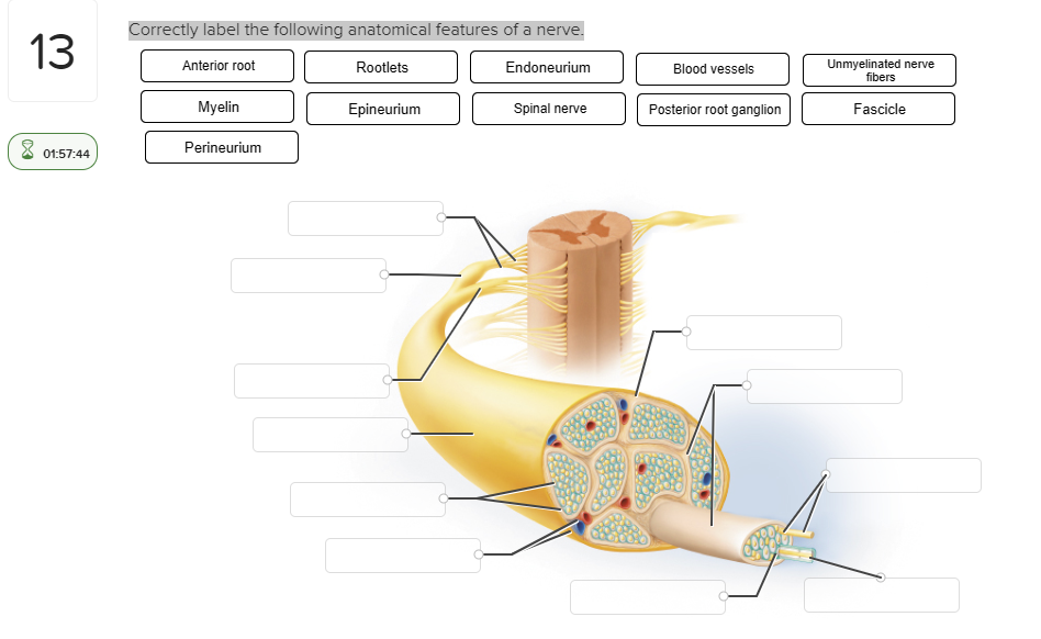
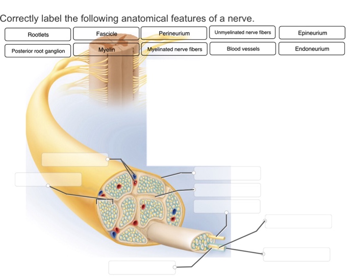
Correctly Label the Following Anatomical Features of a Nerve.
Correctly label the following anatomical features of the spinal cord. 09 ints Nucleus References Axon Myelin sheath Internode Axon terminals Nucleolus Node of Ranvier Dendrites ac Prev 6 of 11 Next pe here to search o Et 2 12 13 14 16 17 710 111 112 A 2 3 4 5 6 7 8 9 0 This problem has been solved.

Anatomy Midterm Lecture Flashcards Quizlet
Stimulation from the __________ nervous system via the __________ nerve causes the secretion of HCl in the stomach.

. All of these parts function to give you the ability to see. The lumbar column has four segments and connects the vertebrae. Controls muscular movement at the subconscious levelPutamen Correctly label the following anatomical features of the cerebellum.
So its important to know the proper labels. In some cases the axons may travel more than one meter. Axons can travel up to a meter depending on the species.
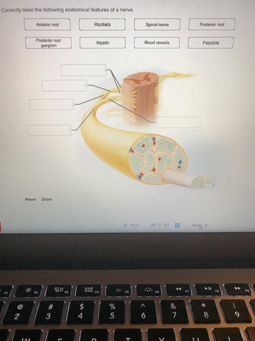
Although most neurons are found in the central nervous system there are also sensory neurons in other areas of the body. The study dealing with the explanations of how an organ works would be an example of _____. Anterior root Spinal nerve Posterior root Posterior root Blood vessels Reset Zoom F6 8 XXXXXXXXXX.
The iris is the inner layer of the eye covering the back two-thirds of the eyeball. Answer Labelled on the left side from top to bottom as 123 and on right side from top to bottom as 4567 1. The cell body dendrites and an axon.
Lateral geniculate nucleus of the thalamus Correctly label the following parts of the brainstem. Anterior median fissure 4. The nerve is also the target of many forms of disease like multiple sclerosis Parkinsons disease and spinal cord injury.
Correctly identify and label the spinal nerves and their plexuses. Identify the spinal nerve plexuses pictured below and drag the innervations to the appropriate category according to which plexus is responsible. Each of the labels below describes a sensory or motor innervation.
The spinal cord is made up of two parts the lumbar column and the brain. Gray commissure White matter Posterior column Meninges Anterior column Lateral column Posterior hom. Which of the following is NOT part of the limbic system.
Es Correctly label the following anatomical features of a nerve. Correctly label the following anatomical features of a nerve. Claustrum caudate nucleus globus pallidus putamen amygdaloid body.
Posterior funiculus it is the white matter of the spinal cord 5. Each nerve has a particular type. The retina the pupil and the lens.
In this article you will learn the anatomical features of the eye including the cornea. It contains nerve cells that carry visual information to the brain. 6 Correctly label the following anatomical features of a neuron.
The optic nerve is the part of the eye that sends electrical signals from the eye to the brain. Correctly label the following anatomical features of a nerve. Correctly identify and label the structures associated with the rami of the spinal nerves.
The autonomic nervous system controls all of the following except the skeletal muscle in the rectus abdominis. Correctly label the following anatomical features of a nerve. See the answer Show transcribed image text.
A typical neuron has three main anatomical features. Primary fissure vermis anterior lobe posterior lobe folia cerebellar hemisphere Label the components of the cerebral nuclei. The lens is the colored part of the iris.
The anatomical features of a nerve can be identified by their function. Axon terminals are axons. The iris is the colored part of the iris.
Gray matter bi Seinal cord and meninges thorack Central canal Spinal nerve Posterior root ganglion Posterior root This problem has been solved. January 9 2022 thanh Lateral funiculus Posterior root of spinal nerve Posterior funiculus Posterior horn Anterior median fissure Spinal nerve Gray commissure Spinal. Most neurons contain a cell body a pair of dendrites and a single axon.
Posterior root of spinal nerve Hope this helps. Expert Answer Transcribed image text. As the brain ages the nerve becomes stretched and distorted.
Posterior horn -it is the sensory horn 2. The eyeball is a sphere containing three primary components. Study Flashcards On Anatomy Physiology Exam 1 Ch.
Most neurons are located in the brain and spinal cord. Label each line on the pressure graph below as representing either the aorta left atrium or left ventricle. Between the iris and the lens is the retinal pigment epithelium.
Correctly label the following anatomical features of a nerve. Therefore correct labels are those that include the dorsal ganglion and the ventral thalamic. The nerve is a tiny bundle of neurons that travels within the body and is responsible for communicating between the brain and the rest of the body.
The following are the main features of the eye. The lumbar segment is the largest of the three. BIOD 152 - Summer 2018.
They also end in small branches called nerve endings. Spinal nerve Blood vessels Fascicle Rootlets Posterior root Myelin ganglion Posterior root Anterior root Previous question Next question. The sensory organs contain the other anatomical components of a nerve.
Correctly label the following anatomical features of the spinal cord. Help manage time and improve learning inside and outside of the lab. The two parts are joined in the center of the spinal column.

A P 1 Final Flashcards Quizlet

Solved Correctly Label The Following Anatomical Features Of Chegg Com

Solved Correctly Label The Following Anatomical Features Of Chegg Com

Solved Correctly Label The Following Anatomical Features Of Chegg Com
No comments for "Correctly Label the Following Anatomical Features of a Nerve."
Post a Comment